

MELANOMA
OVERVIEW
Melanoma is a serious form of skin cancer that develops from melanocytes, the pigment-producing cells of the skin. Although less common than basal cell carcinoma and squamous cell carcinoma, melanoma has a greater potential to spread to lymph nodes and internal organs.
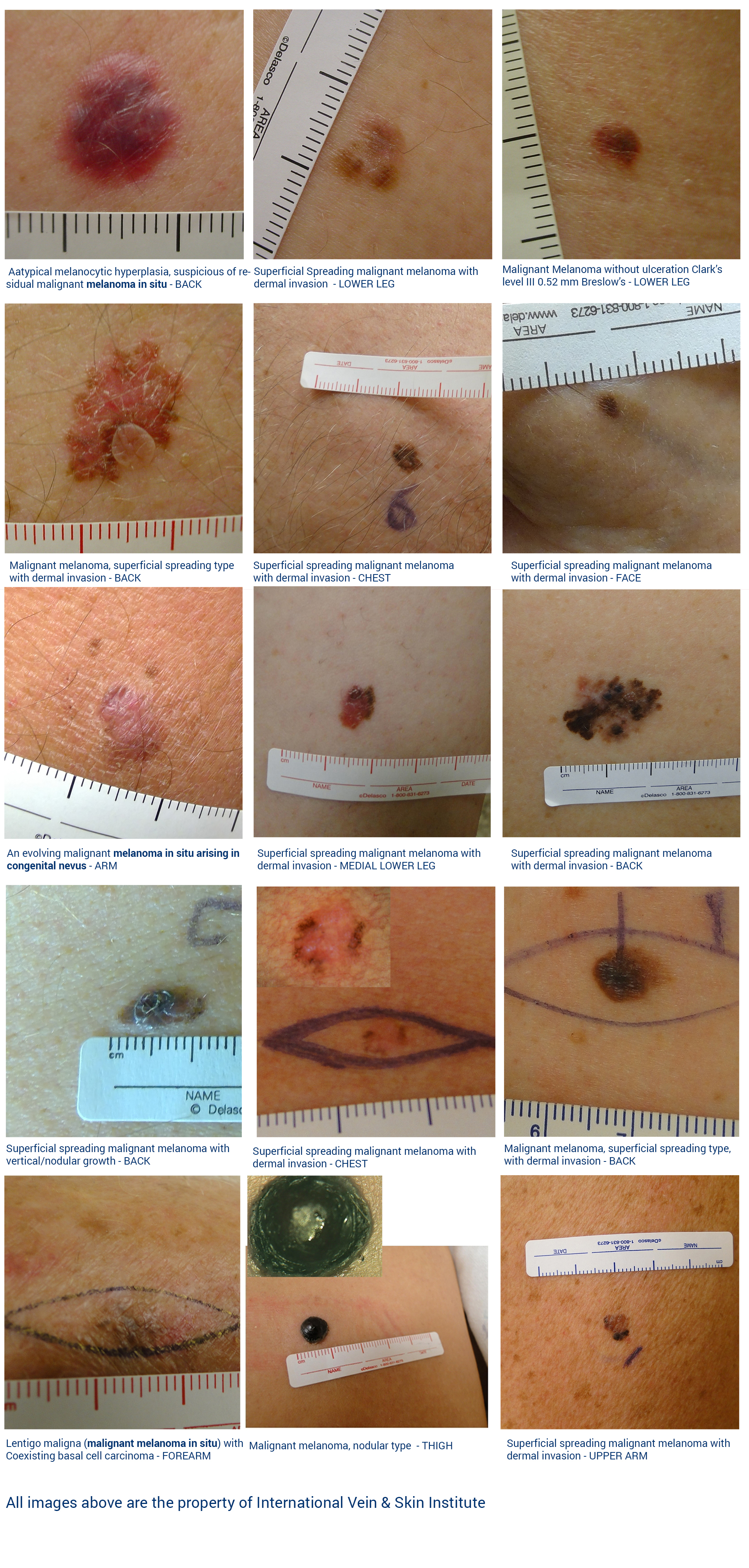
Melanoma can develop anywhere on the body, including areas that receive little or no regular sun exposure. It may arise within an existing mole or develop as a new pigmented or non-pigmented lesion.
Early diagnosis is particularly important because prognosis and treatment are strongly influenced by the depth and stage of the melanoma at the time of diagnosis.
COMMON SIGNS AND APPEARANCE
Melanoma does not always follow a predictable pattern. Warning signs include:
- A new or changing mole
- A lesion that looks different from a person's other moles — the “ugly duckling” sign
- Asymmetry
- Irregular or poorly defined borders
- Uneven or multiple colors
- Increase in size, thickness, or elevation
- New bleeding, crusting, or ulceration
- Itching, tenderness, or another change in sensation
- A new or changing non-pigmented pink or red lesion
A melanoma does not need to demonstrate every classic warning sign to be concerning. Change over time or a lesion that looks distinctly different from surrounding moles deserves medical evaluation.
ABCDE WARNING SIGNS
The ABCDE features provide a useful framework for recognizing changes that deserve closer evaluation:
-
A — Asymmetry
One side looks different from the other. -
B — Border
The edges are irregular, blurred, scalloped, or poorly defined. -
C — Color
Uneven pigmentation or multiple shades of brown, black, red, pink, white, or blue. -
D — Diameter
Larger than 6 mm, or a lesion that is growing regardless of its size. -
E — Evolving
A change in size, shape, color, elevation, surface, symptoms, or behavior.
The ABCDE features are warning signs, not diagnostic requirements. Melanoma can be smaller than 6 mm and can lack one or more of the classic ABCDE characteristics.
RISK FACTORS
Factors associated with increased melanoma risk include:
- History of intense ultraviolet (UV) exposure or blistering sunburns
- Indoor tanning
- Fair skin and tendency to burn easily
- Numerous melanocytic nevi (moles)
- Multiple atypical or dysplastic nevi
- Personal history of melanoma or another skin cancer
- Family history of melanoma
- Immunosuppression
Melanoma can also occur in people without recognized risk factors and in all skin types.
DIAGNOSIS
A lesion suspicious for melanoma cannot be definitively diagnosed by appearance or dermoscopy alone.
A biopsy is performed to obtain tissue for diagnosis. For a lesion clinically suspicious for melanoma, complete excisional biopsy with a narrow margin is generally preferred when feasible, because examination of the entire lesion allows accurate assessment of its histologic characteristics and depth.
The tissue specimen is sent to a pathology laboratory, where it is processed and examined microscopically by a pathologist or dermatopathologist.
The pathology report establishes whether melanoma is present and provides important information used for staging and treatment planning, including the Breslow thickness, ulceration, margin status, and other histopathologic characteristics.
AFTER MELANOMA IS DIAGNOSED
Management after a melanoma diagnosis depends on the pathology findings and stage of disease.
Treatment commonly begins with surgical excision using margins determined by the melanoma's characteristics and depth. Depending on the pathology findings and stage, further management can include:
- Wider surgical excision
- Regional lymph node evaluation, including sentinel lymph node biopsy for appropriate tumors
- Additional staging evaluation
- Referral to surgical oncology, dermatologic surgery, medical oncology, or another appropriate specialist
- Systemic or other oncologic treatment for more advanced disease
The treatment pathway is determined by the individual melanoma diagnosis and staging findings.
👉 Learn more about Next Steps After a Skin Cancer Diagnosis
PROGNOSIS AND FOLLOW-UP
Melanoma prognosis varies substantially according to the stage at diagnosis. Tumor depth, ulceration, lymph node involvement, and distant spread are among the factors that influence prognosis and subsequent management.
Melanoma detected while still localized to the skin generally has a much more favorable prognosis than melanoma that has spread beyond its original site.
Patients diagnosed with melanoma require ongoing skin surveillance, because they have an increased risk of developing additional melanomas and other skin cancers.
WHEN TO SEEK EVALUATION
Medical evaluation is appropriate for:
- A new or changing mole
- A lesion that looks distinctly different from other moles
- A pigmented lesion with asymmetry, irregular borders, or changing color
- A lesion that continues to grow or become elevated
- New bleeding, crusting, ulceration, itching, or tenderness
- Any persistent or changing lesion that causes concern, including a pink or non-pigmented growth
Evaluate a Suspicious Mole or Skin Lesion
A new, changing, unusual, or otherwise concerning mole or skin lesion should be evaluated promptly.
📞 Call (847) 518-9999 to schedule a dermatology evaluation.
Medical Disclaimer
This information is provided for educational purposes only and does not
replace individualized medical evaluation. Treatment recommendations depend
on clinical findings, diagnostic requirements, and patient-specific factors.
Outcomes may vary, and no specific medical result can be guaranteed.
Learn More About