

BASAL CELL CARCINOMA (BCC)
OVERVIEW
Basal cell carcinoma (BCC) is the most common type of skin cancer. It develops from cells within the basal layer of the epidermis and most often occurs on areas exposed to ultraviolet (UV) radiation, particularly the face, scalp, ears, neck, shoulders, and upper trunk.
BCC is generally slow-growing and rarely spreads to distant organs. However, it can continue to enlarge and invade surrounding skin, cartilage, bone, or other structures when left untreated. This is particularly important on the face, nose, eyelids, ears, and other anatomically sensitive areas, where even a relatively small tumor can eventually require more extensive treatment.
Early diagnosis allows treatment while the lesion is generally smaller and more localized.
COMMON SIGNS AND APPEARANCE
Basal cell carcinoma does not always have the same appearance. Depending on its subtype and location, BCC may appear as:
- A pearly, shiny, or translucent bump
- A pink or flesh-colored growth with visible superficial blood vessels
- A persistent red or pink patch
- A lesion that repeatedly bleeds, crusts, or scabs
- A sore that does not heal or repeatedly heals and returns
- A flat, firm, scar-like area
- A slowly enlarging lesion with poorly defined borders
BCC is often painless, allowing some lesions to remain unnoticed or untreated for a long time.
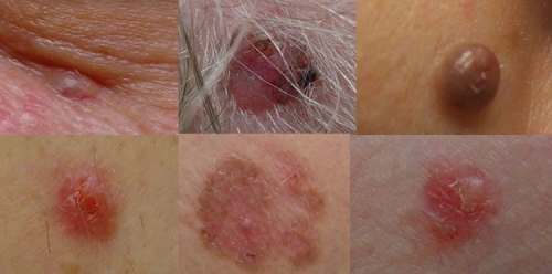
 Images of Basal Cell Carcinoma Lesions
RISK FACTORS
Factors associated with an increased risk of basal cell carcinoma include:
- Cumulative ultraviolet (UV) exposure
- History of significant or repeated sunburns
- Fair skin and tendency to burn easily
- Increasing age
- Personal history of BCC or another skin cancer
- Family history of skin cancer
- Indoor tanning
- Immunosuppression
- Previous radiation exposure
Although BCC occurs more frequently in fair skin, it can develop in people of any skin type.
DIAGNOSIS
A lesion suspicious for basal cell carcinoma cannot be definitively diagnosed by appearance alone.
A biopsy is performed to obtain tissue for diagnosis. Depending on the lesion's size, location, and clinical characteristics, this may involve:
- Shave biopsy
- Punch biopsy
- Excisional biopsy or complete excision
The tissue specimen is sent to a pathology laboratory, where it is processed and examined microscopically by a pathologist or dermatopathologist.
The pathology report confirms the diagnosis and provides information about the histologic subtype and other characteristics important for treatment planning.
AFTER BASAL CELL CARCINOMA IS DIAGNOSED
Treatment planning begins after the pathology diagnosis is established.
Management depends on factors including:
- Histologic subtype
- Tumor size and depth
- Anatomic location
- Whether the tumor is primary or recurrent
- Definition of the clinical borders
- Previous treatment
- Patient-specific risk factors
The goal is to completely treat the cancer while preserving as much healthy tissue, function, and normal anatomy as possible.
For many basal cell carcinomas, treatment is surgical. Certain tumors require specialized techniques or referral based on their location, size, histologic characteristics, or recurrence risk.
👉 Learn more about Next Steps After a Skin Cancer Diagnosis
PROGNOSIS AND FOLLOW-UP
Basal cell carcinoma generally has an excellent prognosis when appropriately treated. Because patients who develop one BCC have an increased likelihood of developing additional skin cancers, continued skin surveillance remains important after treatment.
Previously treated sites should also be monitored for changes that could indicate recurrence.
WHEN TO SEEK EVALUATION
Medical evaluation is appropriate for a skin lesion that:
- Does not heal
- Repeatedly bleeds, crusts, or scabs
- Slowly continues to enlarge
- Develops a pearly, translucent, pink, or scar-like appearance
- Changes after remaining stable for a long period
- Recurs after previous treatment
Evaluate a Suspicious Skin Lesion
A persistent, changing, bleeding, or non-healing skin lesion should be evaluated to establish the diagnosis and determine appropriate care.
📞 Call (847) 518-9999 to schedule a dermatology evaluation.
Medical Disclaimer
This information is provided for educational purposes only and does not
replace individualized medical evaluation. Treatment recommendations depend
on clinical findings, diagnostic requirements, and patient-specific factors.
Outcomes may vary, and no specific medical result can be guaranteed.
Learn More About